|
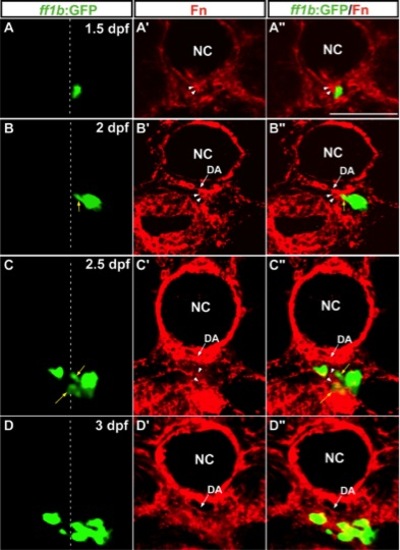
The distribution of Fn around migrating interrenal cells. Interrenal cells (GFP as driven by ff1b promoter in A?D and A′′?D′′) formed a protruding extension at 2 dpf (B, B′′) and dispersed clusters at 2.5 (C, C′′) and 3 dpf (D, D′′), on transverse sections of Tg(ff1bEx2:GFP) embryos. Migrating interrenal cell clusters (denoted by yellow arrows) were closely associated with the Fn protein (red fluorescence in A′?D′ and A′′?D′′; Fn enrichment in the interrenal microenvironment highlighted by white arrowheads) throughout the medial extension process of the interrenal tissue. All sections are oriented with the dorsal side toward the top. White dotted lines indicate the position of the midline. NC, notochord; DA, dorsal aorta. Scale bar?=?50 μM.
|