
Fig. 2
- ID
- ZDB-FIG-130102-13
- Publication
- Kague et al., 2012 - Skeletogenic fate of zebrafish cranial and trunk neural crest
- Other Figures
- All Figure Page
- Back to All Figure Page
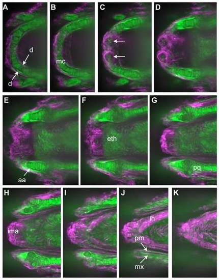
GFP expression persists and reveals pattern of neural crest derivatives in head. A-K represent successive Z-stack projections of five confocal sections each, moving from ventral to dorsal through the head of a 10dpf doubly transgenic embryo. Images have been colored so that green represents GFP+ cells (NC derivatives) and magenta dsRed+ cells (non-NC). Note that throughout the remaining figures, the label associated with NC (GFP or nucCherry) is always shown as green in the two?color overlays. Cartilages known to be NC-derived, including Meckel′s cartilage (B), the ethmoid plate (F), and palatoquadrate (G) are labeled. Also GFP+ are cells in specific areas of ossification, including the dentary (A) and the anguloarticular (E) surrounding Meckel′s cartilage, and the maxilla and premaxilla (J) of the upper jaw. Note also the GFP+ nerve plexus in the lip taste buds (arrows in C), representing their innervation by NC-derived cells of the facial ganglia. Non-NC-derivatives, such as the intermandibularis anterior (ima) and interhyoideus (ih) muscle masses, remain dsRed+. Abbreviations for skeletal structures are listed in Table 3. |
| Genes: | |
|---|---|
| Fish: | |
| Anatomical Terms: | |
| Stage: | Days 7-13 |