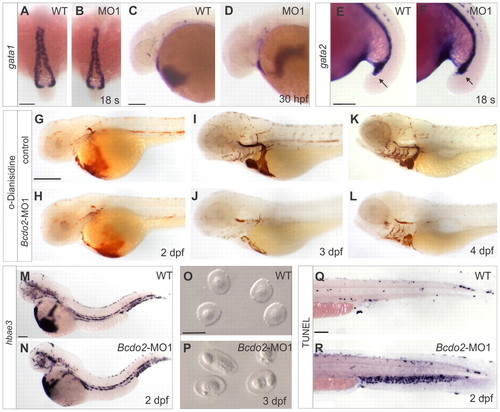
Characterization of the blood phenotype in bcdo2 morphant zebrafish larvae. (A,B) Dorsal views, anterior is upwards. (C-N,Q,R) Lateral views. Anterior is towards the left. (A-F) Whole-mount in situ hybridization for mRNA expression patterns of gata1 and gata2 are similar in morphants and controls. (G-L) o-Dianisidine staining for hemoglobin at different developmental stages of control and Bcdo2 morphant embryos. In 3 dpf bcdo2 morphants (J), staining is reduced when compared with controls (I). (M,N) Staining for hemoglobin (hbae3) mRNA reveals no differences between 2 dpf bcdo2 morphants and controls. (O,P) Blood smears from 3 dpf bcdo2 morphants and control larvae. Blood cells of bcdo2 morphants show fragmented nuclei. (Q,R) TUNEL staining reveals that blood cells of 2 dpf bcdo2 morphants undergo apoptosis. Scale bars: 100 μm in A-F,M,N,Q,R; 250 μm in G-L; 10 μm in O,P. Whole-mount in situ hybridization experiments were conducted with 30 embryos for each condition. o-Dianisidine staining for hemoglobin in morphant and control siblings was conducted with 50 fish per experiment. The experiment was repeated six times with essentially the same outcome.
|
