
Fig. 6
- ID
- ZDB-FIG-120803-34
- Publication
- McCarroll et al., 2012 - Graded levels of Pax2a and Pax8 regulate cell differentiation during sensory placode formation
- Other Figures
- All Figure Page
- Back to All Figure Page
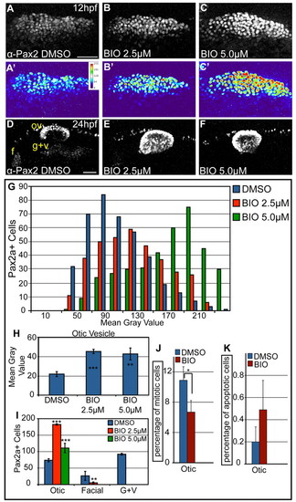
Overactivation of Wnt signaling increases levels of Pax2a expression and biases cells to an otic commitment. (A-C) Confocal projections of DMSO- and BIO-treated zebrafish embryos between 11 and 12 hpf and analyzed for Pax2a expression in the PPA. (A′-C′) Heat maps show increased Pax2a levels after BIO treatment. (D-F) Confocal projection of embryos treated with DMSO or BIO between 11 and 24 hpf and immunolabeled with Pax2a. In BIO-treated embryos, the otic vesicle is larger, whereas EB placodes are significantly reduced (80-100% decrease) (G) Analysis of PPA Pax2a+ cells in control and BIO-treated embryos (2.5 and 5.0 μM) at 12 hpf (n≥354 cells, five embryos per condition) shows dose-dependent increases in expression from low (MGV<120) to high (MGV>120) (χ2, P<<0.001). (H) Comparison of MGVs for Pax2a fluorescence in the otic vesicle showed a significant increase in Pax2a expression levels following BIO treatment (Student′s t-test; **P<0.007; ***P<0.001). (I) Pax2a+ cell number in the otic vesicle showed a 2.47-fold increase in 2.5 μM BIO and a 1.5-fold increase in 5.0 μM BIO versus controls (Student′s t-test; **P<0.01; ***P<0.001). (J) Percentage of Pax2a+ mitotic cells in the otic vesicle dropped at 18 hpf following 10-hour-long BIO treatment (*P<0.05; n=12 embryos per condition). (K) The percentage of Pax2a+ cells that were also Caspase-3+ at 18 hpf in embryos treated with BIO between 10-18 hpf (n=12 embryos per condition) was unchanged. f, facial placode; g+v, glossopharyngeal/vagal placodes; MGV, mean gray value; ov, otic vesicle. Scale bars: 50 μm. |