
Fig. 5
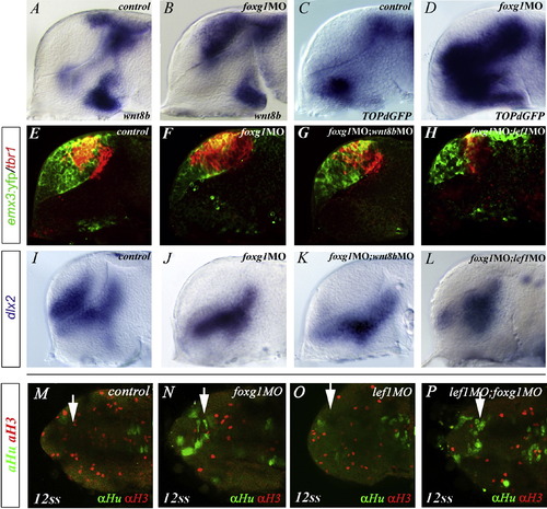
Upregulation of Wnt/β-Catenin Signaling Activity Controls the Expansion of Pallial Fates in foxg1 Morphants(A?L) (A and B) wnt8b and (C and D) topdGFP expression in (A and C) control or (B and D) foxg1MO embryos. Expression of tbr1 (red) and YFP (green) in (E?H) Tg(emx3:YFP) or (I?L) dlx2 , in embryos injected with (F and J) foxg1MO alone or in (G and K) combination with wnt8bMO or (H and L) lef1MO.(M?P) Dorsal views (anterior is oriented toward the left) showing confocal images of double immunostaining of the mitotic marker phosphorylated H3 (red) and the postmitotic neuronal marker HuC (green) in (M) WT, (N) foxg1MO, (O) lef1MO, or (P) foxg1MO;lef1MO double morphants. |
Reprinted from Developmental Cell, 16(4), Danesin, C., Peres, J.N., Johansson, M., Snowden, V., Cording, A., Papalopulu, N., and Houart, C., Integration of telencephalic Wnt and hedgehog signaling center activities by Foxg1, 576-587, Copyright (2009) with permission from Elsevier. Full text @ Dev. Cell