
Fig. 6
- ID
- ZDB-FIG-120601-66
- Publication
- Stevenson et al., 2012 - Hypoxia Disruption of Vertebrate CNS Pathfinding through EphrinB2 Is Rescued by Magnesium
- Other Figures
- All Figure Page
- Back to All Figure Page
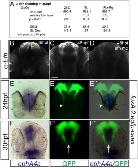
Hypoxia increased ephrinB2a expression in a pattern complementary to its receptor ephA4a. (A) Anti-EfnB2a intensity values at 36 hpf in the telencephalon, either following normoxia, or either hypoxia (1%), or hypoxia with 100 mM magnesium sulfate, from 24–36 hpf. (B) Confocal image of embryo at 36 hpf stained for ephrinB2, demonstrating the region of interest used to calculate α-ephrinB2 intensity. (C, D) Confocal images of embryos at 48 hpf stained for α-ephrinB2, demonstrating that efn morpholino (mo) effectively reduces ephrinB2a expression (D). (E–F3) Whole-mount double immunohistochemistry and in situs for GFP and ephA4a, respectively, in Tg(foxP2-A.2:caax) embryos. (E–E3) During initial axon extension at 24 hpf, axons (arrowhead) travel along the edge of ephA4a expression domain. (F–F3) At 30 hpf, as TCPTc forms (arrow), the axons avoid the area of ephA4a expression. |