Fig. 6
- ID
- ZDB-FIG-120405-56
- Publication
- Eames et al., 2012 - Evolution of the osteoblast: Skeletogenesis in gar and zebrafish
- Other Figures
- All Figure Page
- Back to All Figure Page
|
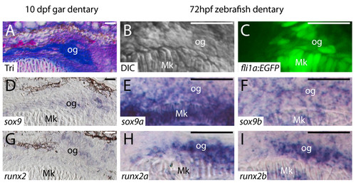
Transcription factor expression in osteogenic condensations of gar and zebrafish is equivalent. A, Trichrome-stained coronal gar section. B, Differential interference contrast image of zebrafish. C, confocal slice of fli1a:EGFP zebrafish. D-I, in situ hybridization on coronal sections. Osteogenic cells of the 10 dpf gar dentary have begun to secrete Aniline blue-stained bone matrix, but do not stain with Alizarin red, as this new matrix is uncalcified (data not shown). Osteogenic cells of the 72 hpf zebrafish dentary are located in an equivalent position. Similar to expression of sox9 in the gar dentary (D), the zebrafish dentary expressed both sox9a (E) and sox9b (F) co-orthologs. Osteogenic cells of the gar dentary expressed transcripts for runx2 (G), and osteogenic cells of the zebrafish dentary expressed both runx2a (H) and runx2b (I) co-orthologs. Scale bars: A-I = 20 μm. Abbreviations: DIC = differential interference contrast; dpf = days post-fertilization; hpf = hours post-fertilization; Mk = Meckel′s; og = osteogenic cells; Tri = Trichrome. |
| Genes: | |
|---|---|
| Fish: | |
| Anatomical Term: | |
| Stage: | Protruding-mouth |

