
Fig. S4
- ID
- ZDB-FIG-120316-50
- Publication
- Sapetto-Rebow et al., 2011 - Maternal topoisomerase II alpha, not topoisomerase II beta, enables embryonic development of zebrafish top2a-/- mutants
- Other Figures
- All Figure Page
- Back to All Figure Page
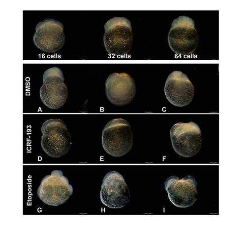
Staging of embryos treated with topoisomerase inhibitors. Top panel: images of normally developed embryos at 16, 32 and 64 cell stages. Bottom panels: example images of "not classified" or malformed embryos treated with 1% DMSO (vehicle control), ICRF-193 or etoposide. This group includes embryos with atypical shape: A) irregular shape, protruding animal pole, B) asymmetric animal pole and uneven cell size, C) irregular shape and opaque, D-E) undefined cell morphology and abnormal transparency, F) small, irregular-shaped animal pole, G) abnormal distribution of cells around yolk, H) undefined cell morphology with abnormal distribution around yolk, I) duplicated/split animal pole. |