
Fig. 6
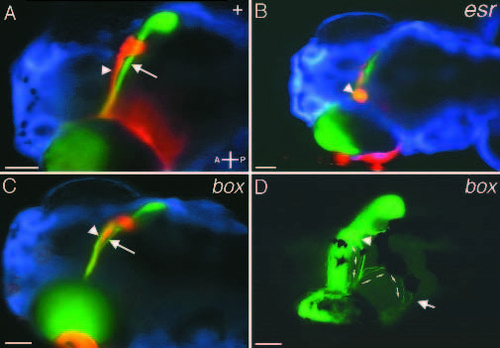
Retinotectal projections in mutants with reduced midline crossing and midline sorting errors. Ventral focal planes. (A) Wildtype projections showing ventral (arrow) and dorsal (arrowhead) brachia of the optic tract. (B) In esrom (and tilsit and tofu, not shown) mutants a large percentage of retinal ganglion cell axons fail to cross the midline. Instead they form an aggregate of fibers (arrowhead) just lateral to the papilla. (C) In boxer mutants, axons from dorsally located retinal ganglion cells do not sort correctly at the optic chiasm. Normally, all axons from dorsal RGCs follow the ventral brachium to the ventral side of the tectum. In boxer (and dackel, not shown), these axons split and follow both the ventral (arrow) brachium and the dorsal (arrowhead) brachium. Axons arriving at the wrong (dorsal) side of the tectum nonetheless find their appropriate ventral target region (see Trowe et al., 1996). (D) In some cases nasal/dorsal RGC axons that grow in the inappropriate (dorsal) brachium fail to enter the contralateral tectal lobe in boxer mutants (arrowhead). These axons (small arrows) project to the ipsilateral tectal lobe by crossing the dorsal midline and terminate in their appropriate retinotopic position (arrow). A, anterior; P, posterior. Scale bars, 100 �m. |
| Fish: | |
|---|---|
| Observed In: | |
| Stage: | Day 5 |