
Fig. S1
- ID
- ZDB-FIG-120202-51
- Publication
- Dohn et al., 2012 - Distinct phases of Wnt/β-catenin signaling direct cardiomyocyte formation in zebrafish
- Other Figures
- All Figure Page
- Back to All Figure Page
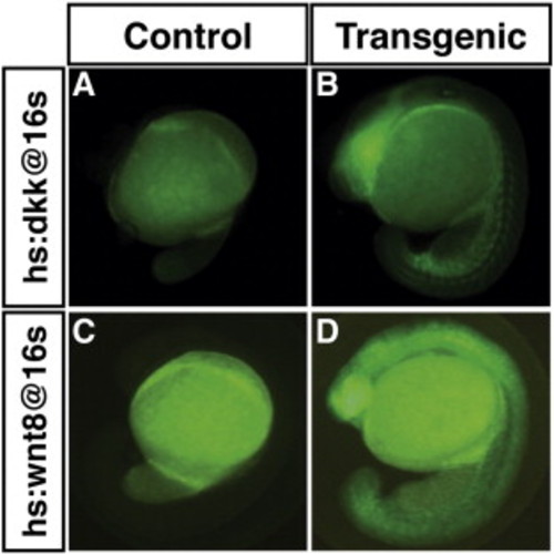
Induction of Dkk-GFP and Wnt8-GFP expression from transgenic lines at the 16 s stage. As previously reported (Stoick-Cooper et al., 2007; Weidinger et al., 2005), both the Dkk and Wnt8 used for the transgenic lines are tagged with GFP facilitating the easy detection of transgenic embryos. (A,C) HCSEs. (B) Representative GFP + Tg(hsp70l:dkk1-GFP)w3 embryo 30 min after the heat-shock. For the Tg(hsp70l:dkk1-GFP)w3 embryos, fluorescence was maximal at ~ 1 h. (D) Representative GFP + Tg(hsp70l:wnt8a-GFP)w34 embryo 2 h after heat-shock. For the Tg(hsp70l:wnt8a-GFP)w34 embryos, fluorescence was maximal at ~ 2 h post-heat shock. For both transgenes, GFP expression could be detected by 30 min post-heat-shock. Overall, relative to each other, the fluorescence induced from the Dkk-GFP transgene is consistently brighter than the Wnt-GFP transgene (data not shown). Equivalent results were obtained with heat-shocks performed at the TB stage. Fluorescence in the yolk in all images is autofluorescence. |
Reprinted from Developmental Biology, 361(2), Dohn, T.E., and Waxman, J.S., Distinct phases of Wnt/β-catenin signaling direct cardiomyocyte formation in zebrafish, 364-76, Copyright (2012) with permission from Elsevier. Full text @ Dev. Biol.