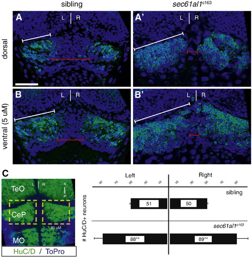
Excess neurons are found in the cerebellum of sec61al1c163 mutants. We quantified neurons in the dorsal-most region of the cerebellum as a comparison to the development of the dorsally-located habenulae. HuC+ neurons are evident in optical sections from the (A) dorsal surface or (B) 5 μm below the dorsal surface of the cerebellar plate (white brackets) at 72 hpf. (A′, B′) More HuC+ cerebellar neurons are evident in sec61al1c163 mutants, particularly in the medial region of the cerebellar plate (red brackets). (C) Representative extended focus image of the hindbrain for orientation; graphs represent data from 5 larvae. Images in A?B′ are single plane images from confocal Z-stacks, with B,B′ taken 5 μm ventral to A,A′, respectively. Image C is an extended focus projection of a confocal Z-stack. Significance determined by Student′s T-test, comparing the left, right, or total cerebellar neurons of sec61al1c163 mutants with siblings (p = 0.0002, 0.002, 0.0005, respectively); n = 5, mutants and siblings. TeO = optic tectum; CeP = cerebellar plate; MO = medulla oblongata. Scale bars = 30 μm.
|
