
Fig. 3
- ID
- ZDB-FIG-111128-30
- Publication
- Wang et al., 2011 - Prdm1a and miR-499 act sequentially to restrict Sox6 activity to the fast-twitch muscle lineage in the zebrafish embryo
- Other Figures
- All Figure Page
- Back to All Figure Page
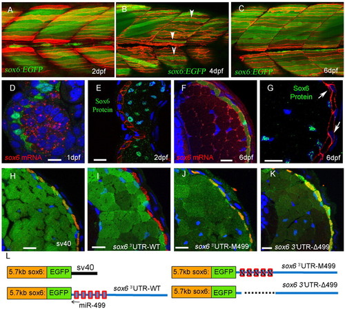
Derepression of sox6 transcription but not translation in slow-twitch fibres. (A-C) Optical sagittal sections of posterior trunk region of Tg(5.7 kb sox6:EGFP)i253; Tg(9.7 kb smyhc1:lyn-tdTomato)i261 zebrafish larvae. At 2 dpf (A), EGFP expression is restricted exclusively to fast-twitch fibres (green) lying beneath the superficial slow-twitch fibres (red). At 4 dpf (B), EGFP expression is detectable in a few slow-twitch fibres (arrowheads). By 6 dpf (C), all slow-twitch fibres express EGFP. (D-G) Transverse cryostat sections of Tg(BACsmyhc1:GFP)i108 (D,F) or Tg(9.7 kb smyhc1:lyn-tdTomato)i261 (E,G) embryos/larvae hybridised with a probe for sox6 mRNA (red) or stained with anti-Sox6 antibody (green), respectively. Arrows indicate slow-twitch fibre nuclei. (H-L) Transverse cryostat sections of 6 dpf larvae carrying the 5.7 kb sox6:EGFP reporter constructs with differing 3′UTRs, as illustrated in L. (H) Expression of EGFP driven by the 5.7 kb reporter construct with the SV40 3′UTR in slow-twitch fibres, as identified by mABF59 staining (red), is repressed by replacement of the SV40 3′UTR with the endogenous sox6 3′UTR (I). Mutation (J) or deletion (K) of the five putative miR-499 target sites restores ectopic EGFP expression in slow-twitch fibres. Note the increased levels of EGFP expression caused by deletion of the miR-499 sites (K). Scale bars: 10 µm. |
| Genes: | |
|---|---|
| Antibodies: | |
| Fish: | |
| Anatomical Terms: | |
| Stage Range: | Prim-5 to Day 6 |