
FIGURE
Fig. 6
Fig. 6
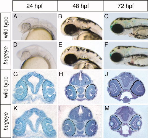
Appearance of head and brain structures in bugeye embryos. Lateral head aspects (A?F) and coronal diencephalic sections (G?M) stained with toluidine blue are shown. No obvious malformations in head or brain anatomy are seen at the indicated time points comparing wild type and bugeye embryos. |
Expression Data
Expression Detail
Antibody Labeling
Phenotype Data
| Fish: | |
|---|---|
| Observed In: | |
| Stage Range: | Prim-5 to Protruding-mouth |
Phenotype Detail
Acknowledgments
This image is the copyrighted work of the attributed author or publisher, and
ZFIN has permission only to display this image to its users.
Additional permissions should be obtained from the applicable author or publisher of the image.
Full text @ Dev. Dyn.