
Fig. 4
- ID
- ZDB-FIG-110922-7
- Publication
- Pyati et al., 2011 - p63 Mediates an Apoptotic Response to Pharmacological and Disease-Related ER Stress in the Developing Epidermis
- Other Figures
- All Figure Page
- Back to All Figure Page
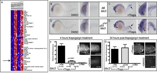
puma Expression Is Increased in a p53-Independent Manner Following Thapsigargin Treatment (A) Microarray analysis of dissected tail tissue revealed increased puma expression following thapsigargin treatment in both AB and p53 mutant embryos. (B?E) Compared to DMSO-treated controls (B), thapsigargin-treated embryos (C) had increased puma expression in the tail (arrow), epiphysis (asterisk), and lens (arrowhead). This increased expression was also observed in thapsigargin-treated p53 mutant embryos (E) compared to DMSO-treated controls (D). (F and G) Knockdown of puma attenuated 4 hr ER stress-induced apoptosis, but not (G) 28 hr ER stress-induced apoptosis. See also Figure S2 and Figure S3. |
| Gene: | |
|---|---|
| Fish: | |
| Condition: | |
| Anatomical Terms: | |
| Stage: | Prim-5 |
Reprinted from Developmental Cell, 21(3), Pyati, U.J., Gjini, E., Carbonneau, S., Lee, J.S., Guo, F., Jette, C.A., Kelsell, D.P., and Look, A.T., p63 Mediates an Apoptotic Response to Pharmacological and Disease-Related ER Stress in the Developing Epidermis, 492-505, Copyright (2011) with permission from Elsevier. Full text @ Dev. Cell