|
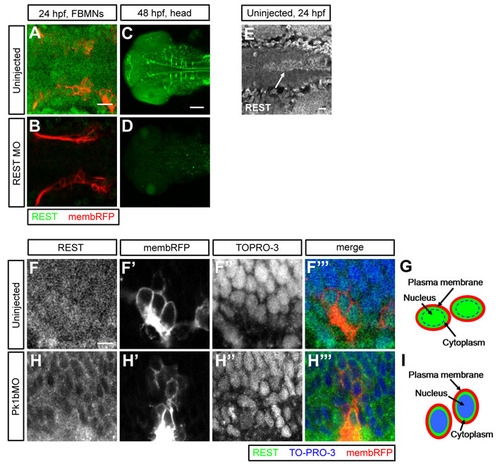
Immunohistochemistry reveals localization of endogenous REST protein. (A-D) Dorsal views of 24 hpf (A,B) and 48 hpf (C,D) Tg(zCREST1:membRFP) embryos. Uninjected (A,C) and REST morphant (B,D) embryos were immunostained for REST (green). FBMNs are visible in A and B (red). REST expression is elevated in several subsets of cells, including reticulospinal neurons (C). Full-length REST protein levels are substantially reduced in REST morphants. E) Dorsal view of the hindbrain in a 24 hpf embryo immunostained for REST. Arrow highlights elevated REST expression in the floor plate. (F-F′′,H-H′′) Single-slice dorsal views of FBMNs (red) in 24 hpf uninjected (F-F′′) and Pk1b morphant (H-H′′) Tg(zCREST1:membRFP) embryos immunostained for REST (green), with nuclei labeled by TO-PRO-3 (blue). (G,I) REST localizes throughout the cell body of wild-type FBMNs, including in the nuclei (G). In Pk1b morphants, REST is reduced in the nuclei and enriched in the cytoplasm (I).
|