
Fig. 4
- ID
- ZDB-FIG-110812-5
- Publication
- Paridaen et al., 2011 - The nucleolar GTP-binding proteins Gnl2 and nucleostemin are required for retinal neurogenesis in developing zebrafish
- Other Figures
- All Figure Page
- Back to All Figure Page
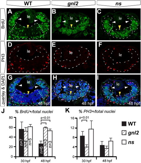
Upon Gnl2 and NS loss of function, retinal progenitors fail to exit the cell cycle in a timely manner. (A–J). Confocal images of BrdU (green) and PH3 (red) double immunolabelling with DAPI overlay (blue) in WT (A, D and G), gnl2 (B, E, H) and ns (C, F and I) embryos at 48 hpf. Retinae are outlined with dashed lines and subdivided into the peripheral (white arrowheads) and central retinae (yellow arrowhead) by dotted lines. In gnl2 (B) and ns (C) embryos at 48 hpf, many BrdU+ cells are present throughout the central retina (yellow arrowheads), whereas in the WT, BrdU+ cells are mainly found at the retinal margin (white arrowheads). The number of PH3+ cells is slightly decreased in gnl2 mutants (E), but not in ns mutants (F). (J) Quantification of the percentage of BrdU+ cells over the total cell number shows that there is no difference at 30 hpf. At 48 hpf, significantly more retinal cells are undergoing S-phase in gnl2 and ns mutants. (K) Quantification of the percentage of PH3+ cells over total cell number shows a reduction in cells undergoing M-phase in gnl2, but not in ns mutants. Lateral is up, anterior to the left. Statistics: Student′s t-test. p-values and experimental numbers indicated in graph. Error bars represent SD. Scalebar 25 μm. |
| Fish: | |
|---|---|
| Observed In: | |
| Stage: | Long-pec |
Reprinted from Developmental Biology, 355(2), Paridaen, J.T., Janson, E., Utami, K.H., Pereboom, T.C., Essers, P.B., van Rooijen, C., Zivkovic, D., and Macinnes, A.W., The nucleolar GTP-binding proteins Gnl2 and nucleostemin are required for retinal neurogenesis in developing zebrafish, 286-301, Copyright (2011) with permission from Elsevier. Full text @ Dev. Biol.