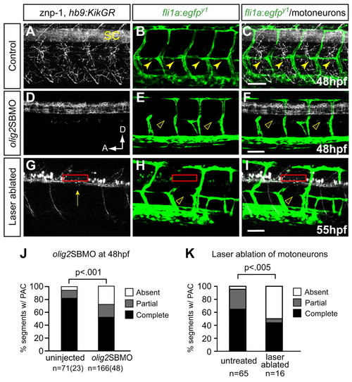
Preventing differentiation or laser ablation of motoneurons prevents PAC formation. (A-F) Lateral views of 48 hpf fli1a:egfpy1 zebrafish embryos either uninjected or injected with olig2 splice-blocking MO (olig2SBMO) showing labeled vessels (green) and neurons (znp-1 antibody, white). Confocal projection. (A) Motor axons grow ventrally from the spinal cord (SC) in controls. (B,C) The PAC forms normally in controls (arrowheads). (D) Motoneurons and their axons are absent in olig2 morphants. (E,F) PAC does not form in olig2 morphants (arrowheads). (G-I) Lateral view of 55 hpf fli1a:egfpy1; hb9:KikGR after photoconversion, with vessels in green and motoneurons in white. Confocal projections. Motoneuron cell bodies were targeted by laser in one somite per embryo (red rectangle), resulting in ablation of motoneurons, (G) missing caudal primary motoneuron (CaP) axon (arrow), and (H,I) missing PAC (arrowhead) in that somite. (J) The PAC was scored in 4-5 somites per embryo in uninjected control embryos (complete, 82�6%; partial, 12�6%; absent, 6�4%) and olig2 morphants (complete, 52�5%; partial, 20�3%; absent, 28�4%). All values are mean�s.e.m.; P-value determined by Mann-Whitney U test. n, number of hemisegments (number of embryos). (K) The number of somites with PACs was quantified in somites that were untreated or successfully laser ablated. Untreated: complete or partial, 95%; absent, 5%. Ablated: complete or partial, 50%; absent, 50%. n, number of hemisegments; P-value determined by Fischer′s exact test. A, anterior; D, dorsal. Scale bars: 50 μm.
|
