
Fig. 6
- ID
- ZDB-FIG-110804-8
- Publication
- Murphy et al., 2011 - A Src-Tks5 Pathway Is Required for Neural Crest Cell Migration during Embryonic Development
- Other Figures
- All Figure Page
- Back to All Figure Page
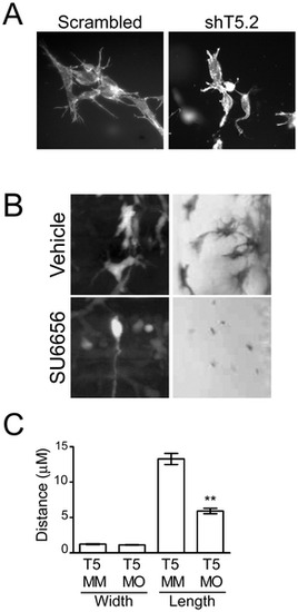
Src- and Tks5-dependent neural crest cell dendritic-like protrusions in 3-D culture and in vivo. (A) Control (scrambled) and Tks5 knocked-down (shT5.2) JOMA1.3 cells were placed in a three-dimensional collagen matrix and cultured for six days. Cells embedded in the collagen matrix were stained for F-actin (phalloidin) and analyzed for differences in cell structure (40�). (B) Neural crest cell and neural crest-derived cell protrusions were qualitatively examined by either enlarging images of neural crest cells in Tg(sox10:RFP) embryos obtained in 4A (left panels) or imaging melanophores in vehicle or SU6656 treated embryos at a higher magnification (23�) (right panels). (C) Control (Tks5 MM injected) and Tks5 morphant Tg(foxd3:GFP) embryos (30 hpf) were fixed and imaged by confocal microscopy. The width and length of protrusions was measured by Volocity software. Mean values (n = 20) and SEM are shown. p values obtained from Student′s t-test. ** denotes p<0.01. (D = dorsal, V = ventral, A = anterior, P = posterior). |