
Fig. S8
- ID
- ZDB-FIG-110516-51
- Publication
- Lazic et al., 2011 - Mef2cb regulates late myocardial cell addition from a second heart field-like population of progenitors in zebrafish
- Other Figures
- All Figure Page
- Back to All Figure Page
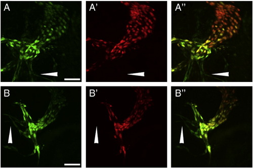
mef2cb did not affect endothelial addition to the arterial pole. Tg(flk1:nlsKikGR) embryos were exposed to UV light at 28 hpf and imaged at 48 hpf to ascertain if endothelial addition at the arterial pole is regulated by mef2cb. Control embryos showed green-only endothelial cells (A, white arrowhead) posterior to the atrial pole that lack red fluorescence (A′). The overlay (A′′) showed endocardial cells to fluoresce green and red. Green-only endothelial cells (compare B to B′) were still present in the mef2cb morphant. Even though there are fewer endocardial cells, reflecting the smaller morphant ventricle, all endocardial cells were green and red (B′′). Green channel (A and B), red channel (A′ and B′), overlay (A′′ and B′′). Scale bar represents 50 μm. |
Reprinted from Developmental Biology, 354(1), Lazic, S., and Scott, I.C., Mef2cb regulates late myocardial cell addition from a second heart field-like population of progenitors in zebrafish, 123-133, Copyright (2011) with permission from Elsevier. Full text @ Dev. Biol.