
Fig. 8
- ID
- ZDB-FIG-110426-1
- Publication
- Schlombs et al., 2003 - Site-1 protease is required for cartilage development in zebrafish
- Other Figures
- All Figure Page
- Back to All Figure Page
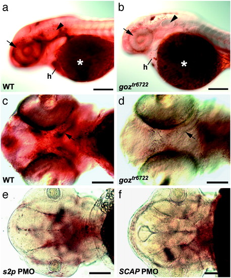
Lipid defects in zebrafish s1p mutants. Oil red O staining of 48 hpf zebrafish larvae. (a and b) Lateral view. (c-f) Ventral view. goz (b and d) and sibling (a and c) larvae and larvae injected with 6 ng of s2p2 (e) and 12 ng of SCAP1 (f) PMOs are shown. Strong lipid staining can be observed around the eye (arrow), the otic vesicle (arrowhead), and in the heart (h) of sibling larvae (a), in addition to the region of developing trabeculae (arrow in c). Both sibling and goz larvae show a strong lipid staining of the yolk (asterisks in a and b). Lipid deposit is severely reduced in all other tissues in goz larvae (b), especially around the eye (arrow) and the otic vesicle (arrowhead). No lipid can be detected around the developing trabeculae in goz (arrow in d). (e and f) Injection of s2p and SCAP PMOs results in the same reduction of lipid. (Scale bars are 200 �m in a and b and 100 �m in c-f.) |
| Fish: | |
|---|---|
| Knockdown Reagents: | |
| Observed In: | |
| Stage: | Long-pec |