
Fig. s2
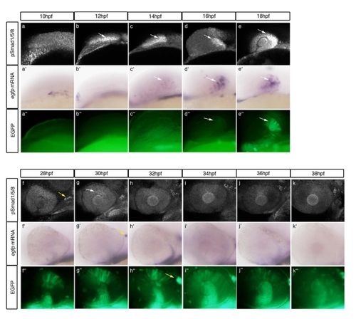
Supporting Information Figure 2. Temporal relationship between pSmad1/5/8 activity and EGFP fluorescence in Tg(bre:egfp) embryos. Embryos were collected every 2 hr between 10?18 hpf and 28?38 hpf and assayed for pSmad1/5/8 via immunofluorescence (a?k), 100�; egfp mRNA via in situ hybridization (a2?k2), 80�; and EGFP fluorescence (a22?k22), 80�. Results demonstrate a 4-hr delay between pSmad1/5/8 expression and EGFP fluorescence, and an 8?10-hr perdurance of EGFP after pSmad1/5/8 is lost. Green indicates bre-driven EGFP expression, 80� magnification. White arrows, dorsal retina; yellow arrows, trigeminal ganglia. All images lateral view, anterior left. |