
Fig. 3
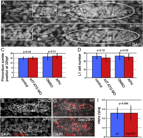
Slowing cell addition does not effect primordium migration, proneuromast deposition or proliferation (A,B) primordia (within white ovals) and proneuromasts (within white rectangles) stained with DAPI (grey) at 32 hpf. (A) Wt primordium. (B) tcf7 morphant primordium. (C) Somite positions of the leading edge of control primordia (blue) and tcf7 morphant or HUA treated primordia (red) at 32 hpf are not significantly different, demonstrating that migration speed is normal in injected embryos. (D). Control and tcf7 morphant or HUA treated primordia deposit proneuromasts that are not significantly different in cell number. TUNEL staining in DMSO treated primordia (E) and HUA treated primordia (F) shows that this treatment does not cause apoptosis within the primordium. (G) BrdU labeling (red) in wt and (H) bap28 mutant primordia, counterstained with DAPI (grey). (I) The BrdU indexes of wt and bap28 mutant primordia are not significantly different. |
Reprinted from Developmental Biology, 349(2), Aman, A., Nguyen, M., and Piotrowski, T., Wnt/?-catenin dependent cell proliferation underlies segmented lateral line morphogenesis, 470-482, Copyright (2011) with permission from Elsevier. Full text @ Dev. Biol.