
Fig. S2
- ID
- ZDB-FIG-110214-63
- Publication
- Maurya et al., 2011 - Integration of Hedgehog and BMP signalling by the engrailed2a gene in the zebrafish myotome
- Other Figures
- All Figure Page
- Back to All Figure Page
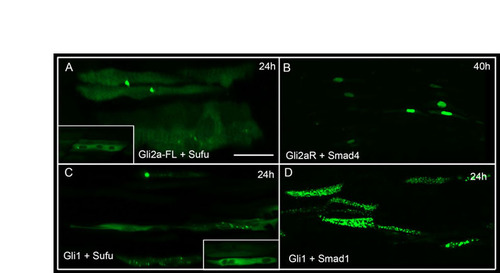
Bi-molecular fluorescence complementation analysis of interacting and non-interacting partners. Parasagittal optical sections of live embryos injected with various constructs expressing Vn- and Vc-tagged proteins from the same UAS promoter and imaged at 24 hours (except in B). (A) Full-length Gli2a and SuFu. Signal is similarly localised to the cytoplasm and excluded from the nucleus. Note punctate accumulations in some fibres. (B) Truncated Gli2a and Smad4 imaged at 40 hours. A few nuclei show signal. (C) Gli1 and SuFu. Signal is localised to the cytoplasm and excluded from the nucleus (inset). (D) Gli1 and Smad1. Note speckled appearance of signal, which shows a cytoplasmic and perinuclear localisation. |