
Fig. 6
- ID
- ZDB-FIG-110112-59
- Publication
- Dolez et al., 2011 - Laminins, via heparan sulfate proteoglycans, participate in zebrafish myotome morphogenesis by modulating the pattern of Bmp responsiveness
- Other Figures
- All Figure Page
- Back to All Figure Page
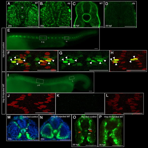
Heparan sulfate proteoglycans are absent in sly embryos and Heparinase III treatment affects Engrailed and pSmad expression in a manner similar to the sly mutant. (A-D) Transverse sections of wild-type (A,C) and sly (B,D) embryos at the 20 s stage (A,B, adaxial cell stacking level) and 26 hpf (C,D, anterior tail level). Dorsal is upwards. In addition to its extracellular signal, anti-HS antibody (in green) labels all nuclei. (E,I) Lateral views of vehicle solution-injected wild-type (E) and Heparinase III-injected wild-type (I) embryos at 26 hpf. Anterior is leftwards, dorsal is upwards. (F-H,J-L) Magnification views (projection of confocal sections) of the regions around somite 10 (F-G,J-K) and somite 18 (H,L) for each type of embryo. (E-L) 4D9 (in green) labels pioneer (arrowheads) and MFF nuclei; Prox1 (in red) labels slow muscle fibre nuclei, including pioneers. Because of the projection, some nuclei artificially overlap. All pioneer nuclei are therefore identified by arrowheads. (E,G,I,K) 4D9 labelling only. (M-P) Transverse sections of vehicle solution-injected wild-type (M,O) and Heparinase III-injected wild-type (N,P) embryos at the 20 s stage (M,N, adaxial cell level) and 26 hpf (O,P, anterior tail level). Dorsal is upwards. (M-P) Nuclear pSmad labelling is in green. (M,N) Nuclei are counterstained with DAPI (in blue). (O,P) 4D9 (in red) labels pioneers. The horizontal white lines delimitate the pSmad-negative central domain. n, notochord; nt, neural tube. Scale bar: 50 μm. |