
Fig. 4
- ID
- ZDB-FIG-101118-47
- Publication
- Hesselson et al., 2009 - Distinct populations of quiescent and proliferative pancreatic β-cells identified by HOTcre mediated labeling
- Other Figures
- All Figure Page
- Back to All Figure Page
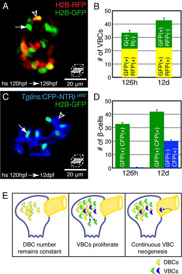
The population of ventral bud derived β-cells (VBCs) expands by proliferation and neogenesis. (A and B) Insulin-HOTcre embryos were injected with H2B-RFP mRNA at the one-cell stage and heat-shocked at 120 hpf. (A) Confocal section of an islet at 126 hpf. (B) Quantification of VBCs per embryo at 126 hpf and 12 dpf, mean ± SEM (Table S1). (C and D) Insulin-HOTcre; Tg(ins:CFP-NTR)s892 embryos heat-shocked at 120 hpf. (C) Confocal section of an islet at 12 dpf. (D) Quantification of β-cells per embryo that had differentiated before the heat-shock (green bars) and those that differentiated after the heat-shock (blue bars), mean ± SEM (Table S1). (A and B). Some cells (DBCs) express both H2B-RFP and H2B-GFP (arrowhead); other cells (VBCs) express only the H2B-GFP label (arrow). The number of VBCs increased between 126 hpf and 12 dpf, reflecting division of existing VBCs. (C and D) β-cells marked by Insulin-HOTcre labeling at 120 hpf express H2B-GFP and CFP (arrow), whereas β-cells that differentiated after the heat-shock only express CFP (arrowhead). β-cell neogenesis contributes a significant proportion of β-cells during larval development (blue bars). (E) β-cells include both DBCs (yellow) and VBCs (green and blue) and the population of VBCs expands through a combination of proliferation (green) and neogenesis from the extra-pancreatic duct (blue), and later, intrapancreatic ducts (data not shown). |