
Fig. S4
- ID
- ZDB-FIG-101117-11
- Publication
- Oka et al., 2009 - The fifth class of Gα proteins
- Other Figures
- All Figure Page
- Back to All Figure Page
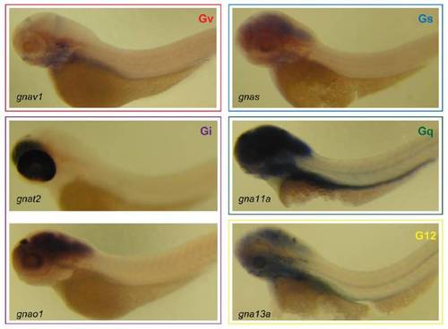
Expression pattern of gnav compared to gna genes of the other 4 classes. Whole-mount in situ hybridization of gnav, gnas, gnat2, gnao1, gna11a, and gna13a probes with 3 dpf zebrafish larvae was performed as described in Fig. S3. All images are lateral views, and anterior is to the left. Note that expression patterns are characteristically different and none from the other 4 classes is similar to that of gnav1. Colored frames enclose genes from the same class (red, Gv; blue, Gs; purple, Gi; green, Gq; yellow, G12). Primers used to clone gna genes are as follows: gnas-fw, 5′-aagactgaggaccagcgaaa-3′; gnas-rv, 5′- gctggacaggctaactggac-3′; gnat2-fw, 5′-ctggtgaagctgccacagta-3′; gnat2-rv, 5′-gcttctctacaagcgccatt-3′; gnao1-fw, 5′-ccagtccaacgctgtctttt-3′; gnao1-rv, 5′- cgctccttgtctccgtactc-3′; gna11a-fw, 5′-cgatcaggttctggtggaat-3′; gna11a-rv, 5′-tgaaaggcgagttggagtct-3′; gna13a-fw, 5′-agaaactgcacatcccttgg-3′; and gna13a-rv, 5′-ttttggctgggcaagtagtc-3′. |