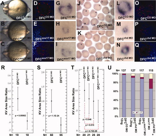
Sox17 and chd are required for proper formation of KV. CO MO (A,D,G: 20 ng; R: 15 ng; J,L,O,S: 2.5 ng; T,U: 12 ng), sox17 MO (B,C,E,F,H,I: 20 ng; R: 15 ng; T,U: 10 ng) or chd MO (K,M,N,P,Q,S: 2.5 ng; T,U: 1.25 ng) were injected into the yolk at the midblastula stage, and embryos were examined at 6?8 somites. Sox17 MO- and chd MO-injected embryos developed small and abnormal KVs as seen in live embryos (A-C; posterior view), by whole mount in situ hybridization with charon (G-N), and by immunostaining for cilia using anti-acetylated tubulin antibody (red) and DAPI (blue) (D-F and O-Q). Scale bars = 0.1 mm in A; 10 �m in D and O. R,S: NIH ImageJ was used to measure the KV area in injected embryos, shown as ratio to the mean control area; error bars are standard deviations; P values are given in the panels. DFCsox17 MO (R) and DFCchd MO (S) embryos showed a significant reduction in KV size compared to controls. T,U: Embryos injected at the midblastula stage with low levels of individual MO (12 ng CO MO, 1.25 ng chd MO, 10 ng sox17 MO) or a combination of 1.25 ng chd MO and 10 ng sox17 MO. KV area was measured as above (T), or lefty2 expression was tested at the -21 somite stage (U).
|
