
Fig. 3
- ID
- ZDB-FIG-100730-5
- Publication
- Eberhart et al., 2006 - Early Hedgehog signaling from neural to oral epithelium organizes anterior craniofacial development
- Other Figures
- All Figure Page
- Back to All Figure Page
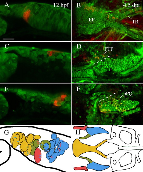
The anterior neurocranium of zebrafish is neural crest-derived. (A,C,E) Lateral views of premigratory crest labeled via photoconversion of Kaede at 12 hpf. (B,D,F) Embryos reimaged at 4.5-5 dpf. (A,B) Cells labeled just posterior to the eye contributed to the ethmoid plate (EP) and trabeculae (TR). (C,D) Neural crest cells slightly more posterior contributed to pterygoid process of the palatoquadrate (PTP). (E,F) Premigratory non-pterygoid palatoquadrate (pPQ) precursors were the most posteriorly localized first arch derivative. (G,H) Schematic representations of fate mapping results show an anteroposterior bias in first arch cartilage elements. Premigratory anterior neurocranium (n=18) and pterygoid process (n=3) precursors (yellow and olive, respectively) are localized more anteriorly than Meckel′s cartilage (MC) (n=1) and palatoquadrate (n=11) precursors (red and blue, respectively). Black outlined circles represent individual labeled embryos. Small colored dots within a field represent secondary cartilage fates from the field of cells. (A,C-G) Lateral views. (B,H) Dorsal views. Anterior is leftwards in all panels. Scale bar: 50 μm. |