
Fig. 3
- ID
- ZDB-FIG-100422-8
- Publication
- Yamamoto et al., 2010 - Two tyrosine hydroxylase genes in vertebrates: New dopaminergic territories revealed in the zebrafish brain
- Other Figures
- All Figure Page
- Back to All Figure Page
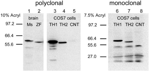
The specificity of polyclonal (left) and monoclonal (right) anti-TH antibodies for TH1 and TH2 proteins was tested on Western blots. Lanes 1 and 2 correspond to proteins extracted from mouse (Ms) and zebrafish (ZF) brains. Lanes 3 and 6 correspond to zebrafish TH1, and lanes 4 and 7 correspond to TH2, both expressed in COS7 cells. The band of the TH2 shows a lower molecular weight than TH1, and it is labeled much weaker than TH1 by the two antibodies. Lanes 5 and 8 correspond to negative controls (CNT), with lane 5 from untransfected COS7 cells, and lane 8 from COS7 cells transfected with a plasmid in which TH2 is inserted in a reverse orientation. The blot has been overexposed, in order to see the TH2 band better. |
| Antibody: | |
|---|---|
| Fish: | |
| Anatomical Term: | |
| Stage: | Adult |
Reprinted from Molecular and cellular neurosciences, 43(4), Yamamoto, K., Ruuskanen, J.O., Wullimann, M.F., and Vernier, P., Two tyrosine hydroxylase genes in vertebrates: New dopaminergic territories revealed in the zebrafish brain, 394-402, Copyright (2010) with permission from Elsevier. Full text @ Mol. Cell Neurosci.