
FIGURE
Fig. S2
- ID
- ZDB-FIG-100422-64
- Publication
- Schonthaler et al., 2010 - The zebrafish mutant bumper shows a hyperproliferation of lens epithelial cells and fibre cell degeneration leading to functional blindness
- Other Figures
- All Figure Page
- Back to All Figure Page
Fig. S2
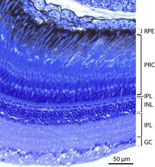
Histological section showing the layers of the adult wild-type zebrafish retina. See Fig. 4I for the retinal morphology in adult bum-/- zebrafish. Abbreviations: GC, retinal ganglion cells; INL, inner nuclear layer; IPL, inner plexiform layer; OPL, outer plexiform layer; PRC, photoreceptor cells; RPE, retinal pigment epithelium. |
Expression Data
Expression Detail
Antibody Labeling
Phenotype Data
Phenotype Detail
Acknowledgments
This image is the copyrighted work of the attributed author or publisher, and
ZFIN has permission only to display this image to its users.
Additional permissions should be obtained from the applicable author or publisher of the image.
Reprinted from Mechanisms of Development, 127(3-4), Schonthaler, H.B., Franz-Odendaal, T.A., Hodel, C., Gehring, I., Geisler, R., Schwarz, H., Neuhauss, S.C., and Dahm, R., The zebrafish mutant bumper shows a hyperproliferation of lens epithelial cells and fibre cell degeneration leading to functional blindness, 203-219, Copyright (2010) with permission from Elsevier. Full text @ Mech. Dev.