|
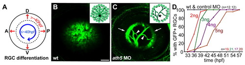
ath5 MO blocks differentiation of early- but not late-born RGCs. (A) Normally, early RGCs differentiate in a wave across central retina (blue arrow); late RGCs are then added centrifugally (red arrows). (B,C) 6 dpf isl2b:GFP, lateral views, anterior towards the left. Insets schematize cell bodies, axons and optic nerve head (star). (B) Wild-type eye shows GFP+ RGCs throughout central retina; axons are obscured by cell bodies. (C) A high dose of ath5MO blocks differentiation of early RGCs, but late RGCs still form (arrows). Without central RGCs, peripheral axons are visible (arrowheads). (D) Dose-response curve showing timing of RGC formation with different doses of ath5MO. In wild type and with 3 ng control MO, GFP+ RGCs appear by 33 hpf. Increasing concentrations of ath5MO increasingly delay the appearance of the first RGCs. A, anterior; D, dorsal; P, posterior; V, ventral. Scale bar: 50 μm.
|