
Fig. 9
- ID
- ZDB-FIG-090721-18
- Publication
- Slanchev et al., 2009 - The epithelial cell adhesion molecule EpCAM is required for epithelial morphogenesis and integrity during zebrafish epiboly and skin development
- Other Figures
- All Figure Page
- Back to All Figure Page
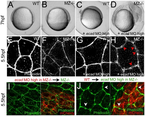
Combined complete loss of EpCAM and Ecad leads to compromised intercellular adhesion within the EVL of early gastrula embryos. (A–D) Live animals at the 70% epiboly stage (7 hpf); dorsal to the right; (E–J) phalloidin stainings of the actin cytoskeleton at 50% epiboly (5.5 hpf). (A,E) wild-type control (WT); (B,F,I) MZepcam mutant control; (C,G) WT injected with high amounts of E-cadherin morpholino oligonucleotide (ecad MO); (D,H,J) MZepcam mutant injected with high amounts of ecad MO. The regular ecad morphant (C) displays an arrest of deep cell epiboly at the equator, as previously described [10]–[12],[87], whereas EVL morphology appears normal (G). In contrast, EVL cells of the ecad;epcam double morphant/mutant embryo lose contact to each other (H; red arrowheads point to sites disassembly sites), and the embryo lyses (D). (I,J) The same EVL disassembly is observed in genetic mosaics, in which ecad;epcam double morphant/mutant EVL are positioned above transplanted wild-type deep cells (J; white arrows to disassembly sites), whereas wild-type EVL above ecad;epcam double morphant/mutant deep cells appear normal (I). Deep cells were transplanted at late blastula stages (3.5 hpf), and immunostained for the tracing marker mCherry in red. |
| Fish: | |
|---|---|
| Knockdown Reagent: | |
| Observed In: | |
| Stage: | 50%-epiboly |