|
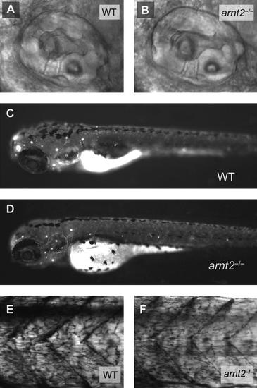
Morphology of the inner ear, neuromasts, and primary motor neurons appears normal in arnt2-/- larvae at 72?148 hpf. Structures of the inner ear, including the semicircular canals and otolith of a WT larva and an arnt2-/- larva at 72 hpf, are similar (A, B). Neuromasts, the lateral line neurons that sense vibration in the water, are stained with DASPEI and appear as white dots on the lateral surface of WT and arnt2-/- larvae (C, D). The DASPEI-stained neuromasts are similar in number and distribution in the two genotypes. ZNP1-stained primary motor neurons are shown in the trunk of a representative WT larva and arnt2-/- larva at 148 hpf (E, F). Skeletal muscle of the trunk is also shown in these lateral views (E, F). Both ZNP1-stained primary motor neurons and trunk skeletal muscle of WT and arnt2-/- larvae appear to be similar.
|

