
Fig. 5
- ID
- ZDB-FIG-090511-19
- Publication
- Willett et al., 1997 - Expression of zebrafish rag genes during early development identifies the thymus
- Other Figures
- All Figure Page
- Back to All Figure Page
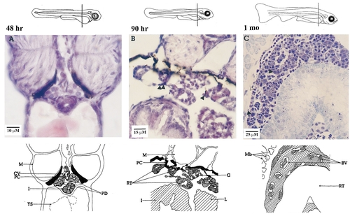
Histological development of zebrafish pronephros. (A) 48 hr, pronephric ducts associated with caudal vein. (B) 90 hr, renal tubules and glomerulae are visible; isolated cells resembling either primitive hematopoietic cells or lymphocyte-like cells appear (▲). (C) 1 month, hematopoietic tissue occupies the space between large renal tubules and blood vessels. The diagram above the photo shows the plane of each section and the sketch below identifies the organs. Photos A and B are of paraffin sections stained with hematoxylin and eosin Y; photo C is of a plastic section stained with toluidine blue. BV, blood vessel; CV, caudal vein; G, glomerlus; I, intestine; L, liver; M, muscle; Mb, muscle bundles; PC, pigment cell; PD, pronephric duct; RT, renal tubule; YS, yolk sac. |
Reprinted from Developmental Biology, 182(2), Willett, C.E., Zapata, A.G., Hopkins, N.A., and Steiner, L.A., Expression of zebrafish rag genes during early development identifies the thymus, 331-341, Copyright (1997) with permission from Elsevier. Full text @ Dev. Biol.