|
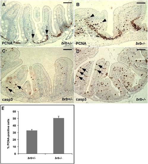
brom bones has increased cell proliferation and apoptosis in the intestinal epithelial cells. (A,B) Representative cross-sections of the anterior intestines from control and mutant fish immunostained for PCNA. Arrows in (A) mark the border of PCNA expression region in the intervillus pocket of a control fish. Arrowheads in (B) indicate PCNA-positive cells in the center region of intestinal epithelium of a brom bones homozygous fish. (C,D) Representative cross-sections of the anterior intestines from control (C) and mutant (D) fish immunostained for active caspase 3. Arrows point to caspase-positive cells. (E) Quantification of the PCNA-positive cell number. Values shown are the percentages of PCNA-positive cells relative to the total number of intestinal epithelial cells per villous cross-section (n = 97 villi from 6 brom bones heterozygous fish and 98 villi from 8 brom bones homozygous fish). Error bars represent s.e.m; P = 2x10⁻⁴ by Student's t test. Scale bar = 50 μm.
|