Fig. 1
- ID
- ZDB-FIG-090114-28
- Publication
- Yelon et al., 1999 - Restricted expression of cardiac myosin genes reveals regulated aspects of heart tube assembly in zebrafish
- Other Figures
- All Figure Page
- Back to All Figure Page
|
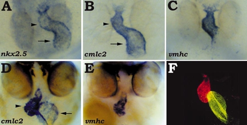
The vmhc gene and S46 antigen are chamber-specific. (A, B, C) Dorsal views of 30-hpf embryos, anterior at the bottom; in situ hybridization showing nkx2.5 (A), cmlc2 (B), and vmhc (C) expression. While nkx2.5 (A) and cmlc2 (B) are expressed throughout both the ventricular (arrowhead) and the atrial (arrow) portions of the heart tube, vmhc (C) expression is restricted to the ventricular portion. (D, E) 48-hpf embryos viewed head-on, dorsal at the top; in situ hybridization with cmlc2 (D) and vmhc (E) riboprobes. As the yolk is gradually absorbed, the ventricle (arrowhead) becomes rostral relative to the atrium (arrow). The expression of cmlc2 (D) is maintained in both chambers, and the expression of vmhc (E) remains restricted to the ventricle. (F) Head-on view of a 48-hpf embryo stained with MF20 (TRITC) and S46 (FITC), dorsal at the top. In this double exposure, red fluorescence indicates MF20 staining of the ventricle, while yellow fluorescence indicates the overlap of S46 and MF20 staining in the atrium. |
Reprinted from Developmental Biology, 214(1), Yelon, D., Horne, S.A., and Stainier, D.Y.R., Restricted expression of cardiac myosin genes reveals regulated aspects of heart tube assembly in zebrafish, 23-37, Copyright (1999) with permission from Elsevier. Full text @ Dev. Biol.

