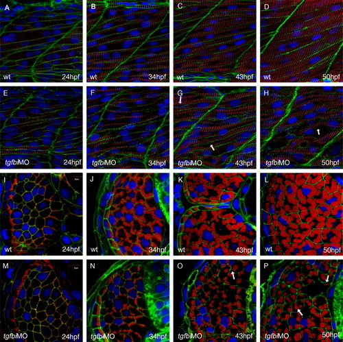
Fibre growth and myofibril bundling is disrupted in tgfbi morphants. Lateral (A-H) and transverse (I-P) aspects of stage matched wild type and morphant embryos. Nuclei are stained blue (DAPI) and the cell membranes are stained green (Lyn-GFP). The red signal reveals the distribution of Z bands (α-actinin) in the lateral views and myofibrils (phallodin) in the transverse sections. In fast fibres of wild type embryos, myofibrillogenesis began along the membrane at 24 hpf (A and I), and muscle fibres continued to grow to become thick myofibres by 50 hpf (D and L). Although myofibrils start to be generated along the sarcolemma in the morphant embryos at 24 hpf (E and M), myofibrillogenesis is subsequently disrupted with myofibrils becoming detached from the sarcolemma (arrows in O and P). The progressive increase in fibre diameter seen in the wild type fast muscle fibres (I-L) is not apparent in the morphant fast muscle fibres (M-P). All images were captured and are reproduced at the same magnification.
|
