|
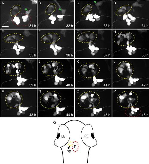
Parapineal cells migrate asymmetrically in flh mutants. All panels are dorsal views of foxd3:gfp labeling in the epithalamus of a single embryo at the times indicated. Parapineal cells are circled in yellow, pineal cells in red; a neural crest cell (which also expresses foxd3:gfp) in A-C is indicated by a green arrowhead. A: The first labeled parapineal cells are apparent near the midline at 31 hr. B-E: They migrate leftward. F-P: They are joined by more leftward-migrating parapineal cells during the subsequent 10 hr. Scale bar = 25 μm. Q: Diagram of a zebrafish larvae, showing the relative position of the pineal (p, red oval) and parapineal (pp, yellow oval) within the head.
|