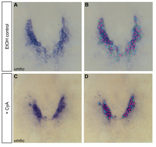
Fig. S1
|
Example of the cell counting technique used to quantify cardiomyocytes at 18-somite and 22-somite stages. (A-D) In situ hybridization detects vmhc expression at the 18-somite stage. Dorsal views, anterior towards the top. In cell counting experiments, the number of cardiomyocytes expressing the gene of interest is determined by marking each cell containing NBT/BCIP precipitate. (A,C) Individual cells are easily identified and quantified as the staining is excluded from the nucleus; nuclei are counted if surrounded by precipitate. (B,D) In these examples, the most intensely stained cells are marked with pink dots, the faintest cells are marked with green dots and the intermediate cells are marked with blue dots. All marked cells are included in the total cell count. |

