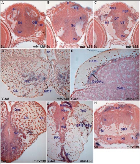
miR-138 expression in the zebrafish brain
miR-138 is expressed in differentiated cells with more restriction compared to miR-124. Expression is largely conserved between larval and adult brain but there are some differences (Tables B,G). With the exception of the medulla oblongata where miR-138 larval expression is widespread and thus difficult to correlate with the adult, we observe the following similarities and differences between larval and young adult expression: miR-138 is expressed in both larval and young adult brains in the olfactory bulb, pallial and subpallial areas, preoptic area, dorsolateral habenular cells, dorsal posterior thalamic nuclei, hypothalamic region, tectal cells, isthmic area and cerebellar cells. Furthermore it is expressed in the adult medial octavolateral nucleus, facial and vagal lobe and reticular formation. This adult hindbrain expression may partially correspond to the larval expression in lateral isthmus and central and medial part of the medulla oblongata. miR-138 is not expressed in the young adult ventral thalamus and semicircular torus whereas it is expressed in these areas at 5dpf. Conversely, it is expressed in adult migrated posterior tuberculum areas, migrated pretectal nuclei, longitudinal torus and cerebellar valvula cells, areas devoid of staining in the larval brain. A. transverse section through the larval telencephalon showing miR-138 expressing cells in the ventral (Sv) and dorsal (Sd) subpallium, pallium (P) and olfactory bulb (OB).
B. oblique transverse section through the larval caudal telencephalon and epithalamus showing miR-138 expressing cells in the preoptic area (Po), dorsal subpallium (Sd), pallium (P) and habenula (Ha).
C. transverse section through the larval diencephalon and optic tectum showing miR-138 expressing cells in the ventral thalamus (VT), dorsal thalamus (DT) and the periventricular gray zone of the optic tectum (pgz).
D. transverse section through the adult olfactory bulb showing miR-138 expressing cells in the internal granular layer (ICL).
E. transverse section through the young adult cerebellum showing miR-138 expressing cells in the cerebellar ganglionic layer (CeGAL).
F. transverse section through the young adult telencephalon showing miR-138 expressing cells in the subpallium (ventral nucleus of the ventral telencephalon, Vv, dorsal nucleus of the ventral telencephalon,Vd) and the pallium (medial zone of the dorsal telencephalic area, Dm).
G. transverse section through the young adult diencephalon showing miR-138 expressing cells in the ventral periventricular zone of the hypothalamus (Hv), dorsal habenular nucleus (Had), parvocellular superficial (PSp) and central (CPN) pretectal nuclei.
H. transverse section through the larval isthmus, hypothalamus and caudal midbrain showing miR-138 expressing cells in the lateral hypothalamic torus (TLa), isthmic area (Is), and cerebellar plate (CeP).
|

