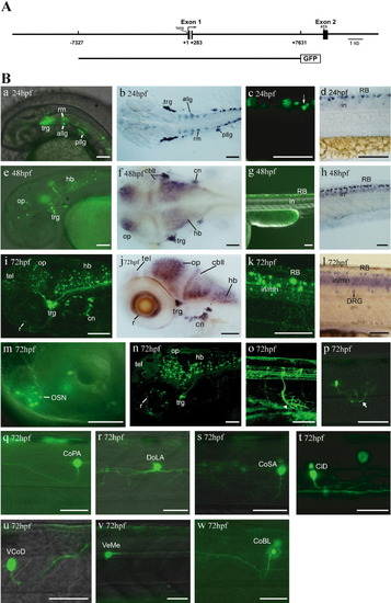
Transient expression profile of scn8aa:GFP. A: The 15-kb fragment of zebrafish scn8aa is cloned into pEGFP-ITR to construct scn8aa:GFP. The gray area in exon 1 (exons are shown in boxes) represents brain-specific regulatory elements shared with mouse SCN8A. B: GFP fluorescence (a, c, e, g, i, k) and in situ hybridization of scn8aa mRNA (b, d, f, h, j, l) in the head and trunk at 24, 48, and 72 hpf. A lateral view is shown, with the anterior to the left and dorsal to the top. m-w: High-magnification views of GFP expression at 72 hpf. Ventral- and dorsal-projecting motoneurons, along with axonal arbors (arrowhead and arrow), are shown in o and p, respectively. GFP-positive interneurons are identified morphologically (q-w). allg, anterior lateral line ganglia; cbll, cerebellum; CiD, circumferential descending interneurons; cn, cranial neurons; CoBL, commissural bifurcating longitudinal interneurons; CoPA, commissural primary ascending interneurons; CoSA, commissural secondary ascending interneurons; DoLA, dorsally longitudinal ascending interneurons; hb, hindbrain; in, interneurons; mn, motoneurons; OP, optic tectum; OSN, olfactory sensory neurons; pllg, posterior lateral line ganglia; r, retina; RB, Rohon-Beard neurons; rm, rhombomere; tel, telecephalon; trg, trigeminal ganglia; VCoD, vental commissural descending interneurons; VeMe, ventral medial interneurons. Scale bars = 100 μm.
|
