Fig. 1
- ID
- ZDB-FIG-080605-32
- Publication
- Kollmar et al., 2001 - Expression and phylogeny of claudins in vertebrate primordia
- Other Figures
- All Figure Page
- Back to All Figure Page
|
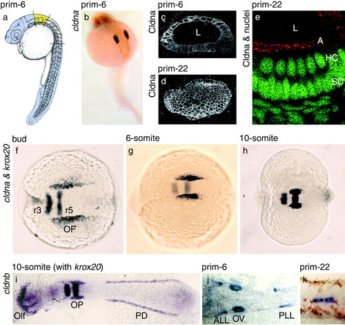
Zebrafish cldna and cldnb mark the earliest developmental stages of the acoustico-lateralis system. (a) Material dissected for the subtractive cDNA hybridization: ear region in yellow; head and tail regions in mauve. [Reprinted with permission from ref. 48 (Copyright 1995, Wiley-Liss, a subsidiary of John Wiley & Sons, Inc.)]. (b and f-h) Whole-mount in situ hybridizations with antisense probes for cldna, which labels the developing ear, and krox20, which marks hindbrain rhombomeres 3 and 5. (c-e) Immunofluorescence detection of Cldna protein throughout the cell membranes of the otic vesicle after acetone permeabilization (c and d), but only at the apical surface of the sensory epithelium, the location of tight junctions, after detergent extraction (e; Cldna in red, nuclei in green). Each panel shows the combination of several adjacent confocal sections; in d, the vesicle was sectioned tangentially. (i-k) Whole-mount in situ hybridizations with antisense probes for cldnb, which labels placodal structures, including those of the ear and lateral-line organ, and for krox20. (k) Migrating placode of the posterior lateral line, with brown pigment cells and boundaries between somites 19 and 22 marked by slanted lines. Views are lateral (a, c-e, and k), dorsolateral (b), or dorsal (f-j), all with anterior to the left, and of whole embryos (a, b, and f-i) or details (c-e, j, and k); in i and j, the specimens have been flattened. L, lumen of the otic vesicle; A, apical surface of the otic vesicle's epithelium; HC, hair-cell nuclei; SC, supporting-cell nuclei; r3 and r5, hindbrain rhombomeres 3 and 5; OF, otic field; Olf, olfactory placode; OP, otic placode; PD, pronephric duct; ALL, anterior lateral-line placode; OV, otic vesicle; PLL, posterior lateral-line placode. |

