
Fig. 6
- ID
- ZDB-FIG-080514-18
- Publication
- Lister et al., 2001 - Duplicate mitf genes in zebrafish: complementary expression and conservation of melanogenic potential
- Other Figures
- All Figure Page
- Back to All Figure Page
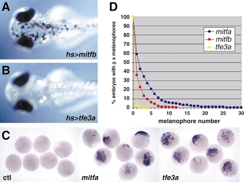
mitfb, but not tfe3a, effectively rescues melanophore development in mitfa mutants. (A, B) Examples of 60-hpf larvae expressing ectopic MiTs under control of the heat shock promoter. mitfb (A) but not tfe3a (B) efficiently restores differentiation of melanophores. A total of 36 out of 143 mitfb-injected embryos had at least one rescued melanophore compared to a single tfe3a-injected embryo with a single melanophore out of 73 injected. (C) Embryos injected with M5-fos:GFP plasmid and the indicated messenger RNA were processed at 8 hpf for in situ hybridization for GFP RNA. mitfa (middle) and tfe3a (right) are each able to transactivate the M-box reporter plasmid in this context. (D) Quantitation of rescue activity by injection of pNP-MiT constructs. Shown on the y-axis are the percentage of injected embryos with an equal or greater number of melanophores as are indicated on the x-axis. mitfa and mitfb both display rescue, but mitfa is somewhat more efficient. Differences between mitfa and mitfb and mitfa and tfe3a are significant as assessed by both the Mann–Whitney U test (P < 0.0001) and Kruskal–Wallis test (P < 0.0001). n = 425 for mitfa, 420 for mitfb, and 379 for tfe3a. No rescue was observed with injection of pNP-mitfa-b692 (n = 235). |
Reprinted from Developmental Biology, 237(2), Lister, J., Close, J., and Raible, D., Duplicate mitf genes in zebrafish: complementary expression and conservation of melanogenic potential, 333-344, Copyright (2001) with permission from Elsevier. Full text @ Dev. Biol.