
Fig. 3
- ID
- ZDB-FIG-080424-55
- Publication
- Unger et al., 2003 - Expression of isotocin-neurophysin mRNA in developing zebrafish
- Other Figures
- All Figure Page
- Back to All Figure Page
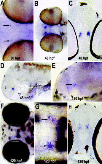
Isotocin-neurophysin (IT-NP) mRNA detected by whole-mount in situ hybridization histochemistry. Anterior is left. (A) Ventral view, IT-NP mRNA is first detected at 36 hpf (black arrows). (B–D) Strong IT-NP mRNA expression is detected in the anterior hypothalamus of the 48 hpf embryo. (B) Ventral; (C) cross section; (D) lateral view. (E–H) Localization of IT-NP mRNA is maintained through 120 hpf of development. (E) Lateral view; (F,G) dorsal views. The outlined area in (F) indicates the region shown enlarged in (G). (H) Cross section. e, epiphysis; hy, hypothalamus; m, midbrain; t, telencephalon; tc, trabecular cartilage. |
| Gene: | |
|---|---|
| Fish: | |
| Anatomical Term: | |
| Stage Range: | Prim-25 to Day 5 |
Reprinted from Gene expression patterns : GEP, 3(1), Unger, J.L. and Glasgow, E., Expression of isotocin-neurophysin mRNA in developing zebrafish, 105-108, Copyright (2003) with permission from Elsevier. Full text @ Gene Expr. Patterns