
Fig. 2
- ID
- ZDB-FIG-080424-17
- Publication
- Whitlock et al., 2003 - Gonadotropin-releasing hormone (gnrh) cells arise from cranial neural crest and adenohypophyseal regions of the neural plate in the zebrafish, Danio rerio
- Other Figures
- All Figure Page
- Back to All Figure Page
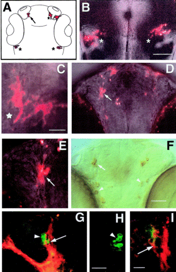
Dil labeling of premigratory cranial neural crest in the living zebrafish embryo. (A) Diagram of 56 h zebrafish head with labeled cells shown in (B–E) colored red: pigment cells (asterisk), cells in trigeminal (star), and cells in terminal nerve (arrow). (B) Dorsal view of 48 h live embryo showing labeled cells in the trigeminal ganglia (asterisks). (A) Ventral view of 56 h live whole-mount embryo head showing two pigment cells (star). (D) Ventral view of 56 h live whole-mount embryo with cells in terminal nerve (arrow). (E) DiI-labeled cells in the terminal nerve (from D) of a live zebrafish embryo (arrow). (F) Anti-GnRH immunoreactivity in TN (arrow) and H population (arrowheads). (G) Ventral view of 56 h whole-mount embryo double labeled for DiI (red) and anti-GnRH antibody (green). DiI is in the GnRH cells of the TN (arrow); some GnRH cells are not double labeled (arrowhead). (H) The contralateral side of this preparation was not labeled for DiI. (I) Higher magnification view of another preparation where the GnRH cells of the TN were double labeled (arrow). Scale bars: (B) 100 μm; (C, E) 25 μm; (D, F) 50 μm; (G, H) 25 μm; (I) 20 μm. |
Reprinted from Developmental Biology, 257(1), Whitlock, K.E., Wolf, C.D., and Boyce, M.L., Gonadotropin-releasing hormone (gnrh) cells arise from cranial neural crest and adenohypophyseal regions of the neural plate in the zebrafish, Danio rerio, 140-152, Copyright (2003) with permission from Elsevier. Full text @ Dev. Biol.