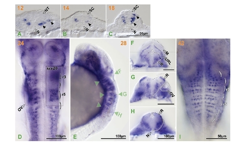
pcdh18 expression in the hindbrain and spinal cord. Orange numbers indicate hours post-fertilization. Black dotted lines outline the structures indicated. Black arrowheads in transverse sections of the trunk of 12–18 hpf embryos (A-C) (dorsal to top) indicate pcdh18-expressing cells in the lateral neural tube and spinal cord. (D) Dorsal view of a 24 hpf embryo stained for pchd18 and krx-20 with single-color double in situ hybridization. Parentheses indicate rhombomeres. Inset shows a focal plane of an area of krx-20 expression which is located ventral to that of pcdh18 expression. Green arrowheads in the lateral view of a 28-hpf embryo (E) indicate focal planes in hand-cut sections (F-H)(dorsal to top) made from the very same embryo. (F) A composite picture of two closely adjacent focal planes. (I) A dorsal view of the hindbrain (rostral to the top) of a 42 hpf embryo with patterns of transverse dotted lines. Abbreviations: M, midbrain; N, notochord; NT, neural tube; OV, otic vesicle; R, rhombencephalon; r3 & 5, rhombomere 3 & 5; SC, spinal cord; URL, upper rhombic lip.
|
