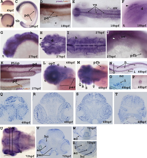
In situ localization of ptmab at indicated stages. A-C,G,L: Lateral view of embryo. E,H,I,M,U: Dorsal view of embryo. K,N: Tail region of the embryo. Double in situ hybridizations for ptmaa and for cdh17 (D, inset in K). F: Higher magnification of the head region. J: Detail of the trunk region of the embryo. O: Detail of the transverse section indicated by the black line in N. P: Magnification of a neuromast). Q-T: Transverse sections indicated by the black lines in M. V,W,Z: Transverse and longitudinal sections indicated by the black lines in U. White arrowhead, the anterior lateral line placode; black arrowhead, the olfactory placode. ac, amacrine cells; c, cerebellum; d, diencephalon; ec, ectoderm; h, hindbrain; hc, horizontal cells; i, intestine; l, lens; n, neuromasts; nc, notochord; opT, optic tectum; ov, otic vesicle; par, pharyngeal arches region; pd, pronephric ducts; pfb, pectoral fin bud; PLLp, posterior lateral line precursor; t, telencephalon; tb, tailbud; vz, ventricular zone.
|
