
Fig. 4
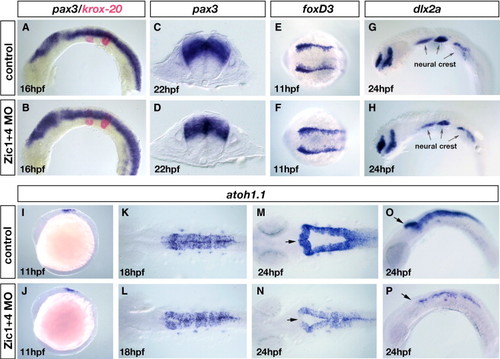
Dorsal fates are selectively altered in Zic1 and Zic4 morphants without affecting early dorsal?ventral (DV) or anterior?posterior (AP) patterning. (A?D) RNA in situ hybridization with a DV marker pax3 (blue in panels A?D) and an AP marker krox20 (red in panels A, B). Lateral view of 16 hpf embryos (A, B) and transverse sections at the level of r5 of 22 hpf embryos (C, D). DV and AP marker distribution and expression are established normally in Zic1 + 4 morphants (B, D) compared to controls (A, C). (E?H) Dorsal view of 11 hpf embryos labeled with the early neural crest induction/specification marker foxD3 (E, F) and lateral view of 24 hpf embryos labeled with a neural crest migration marker dlx2a (G, H). Arrows in panels G and H indicate the expression of dlx2a in the pharyngeal arches. Neural crest induction and migration is unaltered in Zic1 + 4 morphants (F, H) compared to controls (E, G). (I?P) atoh1.1 expression labels dorsal hindbrain progenitors, stages as indicated: lateral views (I, J, O, P), and dorsal views (K?N). At 11 hpf atoh1.1 expression is unaffected in Zic1 + 4 morphants (J) compared to controls (I). At 18 hpf expression is slightly reduced in Zic1 + 4 morphants (L) compared to controls (K). By 24 hpf atoh1.1 expression levels and domain size are dramatically reduced in Zic1 + 4 morphants (N, P) compared to controls (M, O). Arrows in panels M?P indicate the anterior region of r1, where expression of atoh1.1 is lost in Zic1 + 4 morphants. |
| Genes: | |
|---|---|
| Fish: | |
| Knockdown Reagents: | |
| Anatomical Terms: | |
| Stage Range: | 1-4 somites to Prim-5 |
Reprinted from Developmental Biology, 314(2), Elsen, G.E., Choi, L.Y., Millen, K.J., Grinblat, Y., and Prince, V.E., Zic1 and Zic4 regulate zebrafish roof plate specification and hindbrain ventricle morphogenesis, 376-392, Copyright (2008) with permission from Elsevier. Full text @ Dev. Biol.