
Fig. 7
- ID
- ZDB-FIG-070822-68
- Publication
- Schoenebeck et al., 2007 - Vessel and blood specification override cardiac potential in anterior mesoderm
- Other Figures
- All Figure Page
- Back to All Figure Page
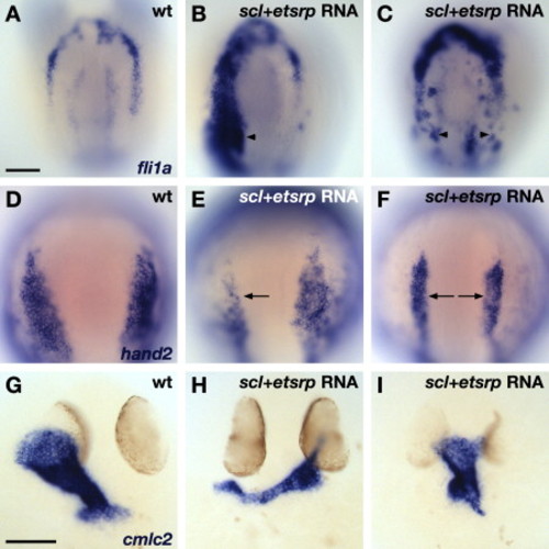
Overexpression of scl and etsrp Promotes Vasculogenesis and Inhibits Heart Formation. In situ hybridization depicts expression of fli1a (A–C), hand2 (D–F), and cmlc2 (G–I) in wild-type (A, D, and G) and scl+etsrp RNA-injected embryos displaying severe (B, E, and H) and moderate (C, F, and I) phenotypes. Dorsal views, anterior to the top, at the 7 somite (A–F) and 30 hpf (G–I) stages. The scale bars represent 100 μm. (A–C) Overexpression of scl and etsrp induces ectopic expression of fli1a, including expression within the HFR (arrowheads; severe phenotype, n = 7/15; moderate phenotype, n = 5/15). (D–F) RNA-injected embryos exhibit reduced expression of hand2 (arrows; severe phenotype, n = 14/34; moderate phenotype, n = 15/34). (G–I) RNA-injected embryos exhibit small, dysmorphic hearts (severe phenotype, n = 9/11; moderate phenotype, n = 2/11). |
Reprinted from Developmental Cell, 13(2), Schoenebeck, J.J., Keegan, B.R., and Yelon, D., Vessel and blood specification override cardiac potential in anterior mesoderm, 254-267, Copyright (2007) with permission from Elsevier. Full text @ Dev. Cell