
Fig. 6
- ID
- ZDB-FIG-070815-24
- Publication
- Zhao et al., 2007 - Genetic defects of pronephric cilia in zebrafish
- Other Figures
- All Figure Page
- Back to All Figure Page
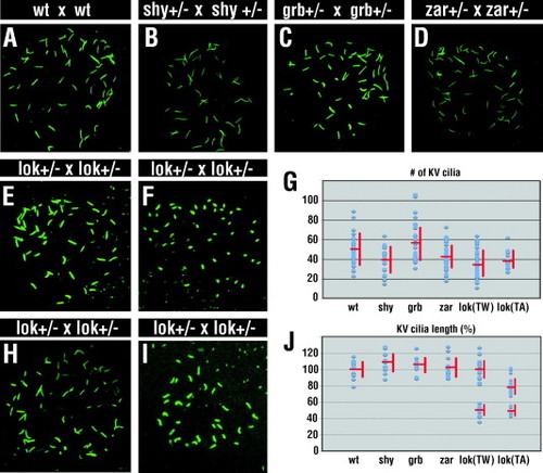
Phenotype of Kupffer?s vesicle. Kupffer?s vesicle (KV) cilia visualized with anti-acetylated tubulin antibody at the 10-somite stage. (A) KV cilia of a typical embryo collected from a control cross between wild-type animals. (B?D) KV cilia of embryos from crosses between two mutant heterozygotes of shy (B), grb (C), and zar (D). Individuals with roughly the average number of cilia are shown. (E, F, H, and I) Phenotypes of KV cilia in two typical embryos collected from a cross between lokto237b TU/WIK heterozygotes (E,F) or lokto237b TU/AB heterozygotes (H,I). In comparison to presumptive wild-type individuals from the same genetic background (E,H), lokto237b mutant embryos display shorter KV cilia (F,I). (G) The number of KV cilia in wild-type and mutant embryos. (J) A graph of KV cilia length in mutant lines relative to the wild type, which is set to 100%. Each dot represents the average length of at least 9 cilia in one embryo. Horizontal and vertical bars in (G,J) indicate the average and the standard deviation respectively. As length measurements in (J) appear to form two clusters, which presumably represent homozygous mutant and phenotypically wild-type embryos, the average and the standard deviation are provided for each cluster separately. TW, Tuebingen/WIK genetic background; TA, Tuebingen/AB genetic background. Embryos in panels (A?D) were of homozygous Tuebingen background. |
| Fish: | |
|---|---|
| Observed In: | |
| Stage: | 10-13 somites |
Reprinted from Mechanisms of Development, 124(7-8), Zhao, C., and Malicki, J., Genetic defects of pronephric cilia in zebrafish, 605-616, Copyright (2007) with permission from Elsevier. Full text @ Mech. Dev.